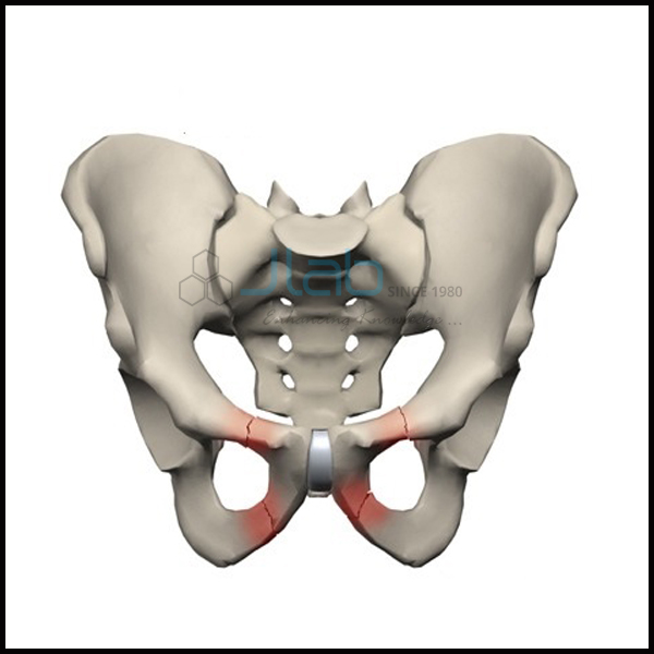

Tethered cord syndrome is a disorder that occurs when tissue attached to the spinal cord limits its movements. It consists of five separate vertebrae that fuse during adulthood. The spinal cord normally hangs freely within the spinal canal. The sacrum is a single bone located at the base of your spine. In most cases, spina bifida occulta causes no symptoms and doesn't need treatment. Spine MRI scan to determine soft tissue damage to the ligaments and discs, and assess spinal cord injury X-rays to show the alignment of the bones along your. A very mild form of this condition, called spina bifida occulta, occurs when the spine doesn't close properly around the spinal cord, but the cord remains within the spinal canal. The spinal column ends above the sacrum but the nerves continue down and through the sacral bone in the form of the. Rarely, sacral dimples are associated with a serious underlying abnormality of the spine or spinal cord. It is a congenital condition, meaning it's present at birth. There are no known causes for a sacral dimple. It's usually located just above the crease between the buttocks. Sacrum: This triangle-shaped bone connects to the hips.
Sacral spine skin#
Most sacral dimples are small and shallow.Ī sacral dimple is an indentation or pit in the skin on the lower back. The lumbar spine bends inward to create a C-shaped lordotic curve. The goal of surgery for a sacral spine cancer is to remove all of the cancer and some of the healthy tissue that surrounds it.

It can also be used to monitor the progression of a disease like osteoporosis or to determine if a treatment you’re having is working.A sacral dimple is an indentation or pit in the skin on the lower back - usually just above the crease between the buttocks. Treatment for chordoma in the sacral spine If the chordoma affects the lower portion of the spine (sacrum), treatment options may include: Surgery. It transmits the total body weight between the lower appendicular skeleton and the axial skeleton. It can be used to view an injury from a fall or accident. The sacrum is the penultimate segment of the vertebral column and also forms the posterior part of the bony pelvis. Your doctor could order a lumbar spine X-ray for a variety of reasons. The lumbosacral joint is the site of most movements of the lumbar spine. coccyx 2) The is a basin-shaped structure that supports the spinal column and protects the sacral plexus. According to the Mayo Clinic, a lumbar spine X-ray can show whether you have arthritis or broken bones in your back, but it can’t show other problems with your muscles, nerves, or disks. When focusing on the lower spine, an X-ray can help detect abnormalities, injuries, or diseases of the bones in that specific area.

The lumbar spine also has:Īn X-ray uses small amounts of radiation to view your body’s bones. The thoracic spine sits on top of the lumbar spine. The coccyx, or tailbone, is located below the sacrum. The sacrum is the bony “shield” at the back of your pelvis.

The sacrum and the iliac bones (ileum) are held together by a collection of strong ligaments. The lumbar spine is made up of five vertebral bones. As a result, the SI joints connect the spine to the pelvis. This is the spinal groove formed by the transverse process of that vertebra. Now glide gently to one side of this and you will feel a dip. Place your finger or thumb on your subject’s spinous process. The function of the sacral vertebrae is to. A lumbosacral spine X-ray, or lumbar spine X-ray, is an imaging test that helps your doctor view the anatomy of your lower back. The spinous process is the most central bony protrusion. sacral vertebrae The vertebrae that lie between the lumbar and the caudal vertebrae in the vertebral column.


 0 kommentar(er)
